Meet the dynamic duo scanning every autism mouse brain
At the Mouse Imaging Centre in Toronto, Jacob Ellegood and Jason Lerch are taking on autism’s complexity by scanning the brain of every autism mouse model they can acquire.
Comments
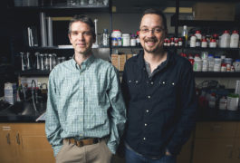
At the Mouse Imaging Centre in Toronto, Jacob Ellegood and Jason Lerch are taking on autism’s complexity by scanning the brain of every autism mouse model they can acquire.
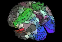
Researchers have charted the human cerebral cortex in unprecedented detail, adding to what is known about the brain’s bumpy outer layer.
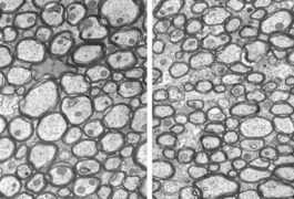
Unusually thin nerve fibers in the brain may underlie the motor difficulties seen in children with Angelman syndrome, an autism-related condition.

Techniques used in behavioral interventions could help scientists scan the brains of children who have both autism and intellectual disability.
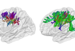
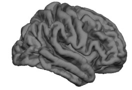
An intricately pleated brain may underlie the highly organized connections between nearby neurons in people with autism.
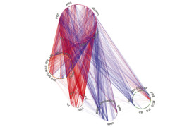
A new set of charts shows how brain connections can change as children grow, and can serve as a reference for detecting problems with attention.
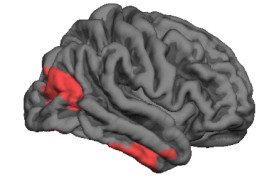
A brain region involved in reading faces has fewer folds in toddler boys with autism than it does in controls, a structural difference that could be related to social difficulties.
A brain imaging technique called magnetoencephalography characterizes not just what is happening in the brain, but also where and when, making it ideally suited for studying autism.
Certain regions of the brain’s bumpy shell become unusually thick and convoluted over time in children with autism.
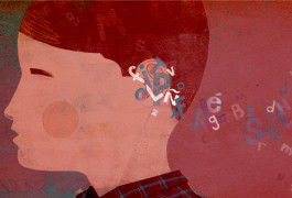
The brain’s sound-processing machinery may mature slowly in children with autism.