THIS ARTICLE IS MORE THAN FIVE YEARS OLD
This article is more than five years old. Autism research — and science in general — is constantly evolving, so older articles may contain information or theories that have been reevaluated since their original publication date.
Two known features of the autism brain — dense connections between nearby neurons and excess folding of the brain’s surface — may be linked, according to a new study1. The findings suggest that excess surface pleating underlies the unusually strong local connections in the brains of people with autism.
Linking the mapping of connections to brain folds — which researchers can directly measure — helps ground this mapping in a physical reality, says lead researcher Christine Ecker, professor of child and adolescent psychiatry at the Goethe University in Frankfurt, Germany. “The way the brain is folded tells us a little more about its connections,” she says.
Neurons consist of cell bodies called gray matter and long projections that connect them, called white matter. By measuring how easily water diffuses across these white-matter fibers, researchers can infer how organized the neuronal fibers that connect brain regions are and, by proxy, how connected these regions are.
By tracing these tracts, researchers have shown that the brains of people with autism may have too many connections between nearby neurons, and too few between those spanning long distances. Studies of functional connectivity — which measure how synchronized different regions are in their activity — support these results.
Studies have also indicated that the brains of some people with autism have unusual patterns of folds in the cerebral cortex, the brain’s outer shell. The findings have been inconsistent, however. Some teams report especially convoluted brain surfaces in certain brain regions in autism, whereas others show the opposite in other regions.
The latest study, published in April in Cerebral Cortex, is the first to rigorously combine two different brain imaging approaches to investigate a possible link between surface folds and the neural connections within.
This type of ‘multimodal imaging’ is a powerful way to gain insights into the autism brain, says Roger Jou, assistant clinical professor at the Yale Child Study Center, who was not involved in the study. But researchers rarely combine data from different brain imaging measures because it’s technically difficult. “It’s nice to see high-quality papers that are actually doing it,” he says.
Double take:
Ecker and her colleagues scanned the brains of 51 men with autism and 48 controls using magnetic resonance imaging to produce a three-dimensional picture of brain folding. Instead of analyzing only regions implicated in autism, the researchers calculated the degree of folding at 300,000 points across the surface of the brain.
They determined that the participants with autism have more folds than controls do in a cluster of regions on the left side of the brain, including the motor cortex. They found that the surface area of this part of the brain is the same in both groups, suggesting that the tissue is simply more tightly folded in the autism brains.
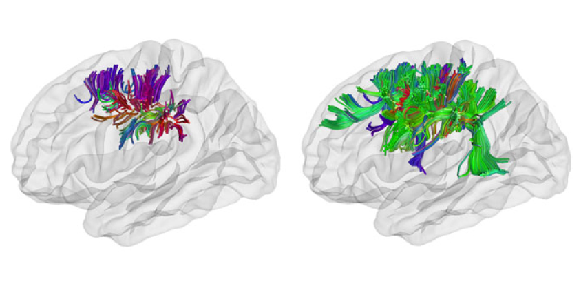
The researchers then used diffusion tensor imaging, the water diffusion technique, to assess the integrity of neuronal tracts that go to or from the region with altered folding. They found that short tracts in this region are more organized in autism brains than in controls, which is consistent with previous results. They saw no difference in long tracts.
The greater the integrity of the tracts, or ‘connectivity,’ the more folds a brain has. This association holds for all of the participants, regardless of whether they have autism. People with autism are more starkly different from controls in terms of brain folding than they are in terms of connectivity, however.
Scratching the surface:
This discrepancy hints that changes in folding are the primary problem in autism, and that differences in the nerve fibers follow from the gray-matter alterations, Ecker says. “We’re basically saying that autism doesn’t affect white matter directly,” she says.
To confirm this thinking, researchers would need to examine brains over time to determine which comes first: differences in connections or in folding.
“So far, we’ve only looked in adults, so we can’t really answer questions of causality,” Ecker says. “We’d really love to look at a longitudinal sample of really young infants to see how that correlation between gray and white matter develops over time.”
The new study may only be scratching the surface of changes in autism brains, says Pratik Mukherjee, professor of radiology at the University of California, San Francisco, who was not involved in the study.
The left-side regions the study highlights have no clear links to autism, he says, but the study may have too few participants to detect more relevant regions, such as those in the so-called social brain network. The number of participants is good for an imaging study, but possibly too low to account for the variation in autism among people — which could mask effects in other regions, Mukherjee says. “What they’re finding is only a very partial effect,” he says.
By joining the discussion, you agree to our privacy policy.