THIS ARTICLE IS MORE THAN FIVE YEARS OLD
This article is more than five years old. Autism research — and science in general — is constantly evolving, so older articles may contain information or theories that have been reevaluated since their original publication date.
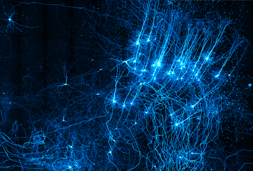
A microscope that sections brain tissue as it scans can trace the tangled paths of thousands of neurons through the entire brain. The device, described yesterday at the 2015 Society for Neuroscience annual meeting in Chicago, could illuminate brain circuits altered in autism.
Traditional methods for tracing neurons involve labeling a neuron, slicing the brain into thin sections and then tracing out the pieces of the neuron in each slice.
“With a lot of work, you can see maybe 10 or 20 neurons,” says Michael Economo, research scientist at the Howard Hughes Medical Institute Janelia Research Campus in Ashburn, Virginia. “We’re trying to find ways to scale that up so you can see hundreds or thousands.”
To scale up and automate the tracing process, Economo and his colleagues developed a microscope with a built-in vibratome — a machine that carefully cuts ultra-thin sections of brain tissue. Instead of scanning sections of brain tissue one at a time, the microscope starts with the whole brain and progressively slices cross-sections of the tissue from top to bottom.
To test the microscope, the researchers first injected a rabies virus with a fluorescent tag that labels neurons into a mouse’s brain. One week later, they removed the brain and submerged it in a clearing solution to make the tissue transparent. They then embedded it in gelatin and placed it on the microscope stage.
Brain building:
The microscope scans a three-dimensional “slab” of brain roughly 250 micrometers thick, Economo says. It then cuts off a slightly thinner section, between 100 and 200 micrometers thick. This process repeats about 20,000 times until the entire brain has been scanned — which takes about a week.
By scanning a thick piece of brain and then removing only part of it, the microscope captures some of the same tissue in two subsequent scans. This allows it to line up features from one image to the next and blend them into a detailed reconstruction of neurons winding their way through the brain.
“It covers every micron of the brain in all three dimensions at very high resolution,” Economo says.
Because the rabies virus can jump across the gaps between neurons, called synapses, the researchers could visualize the connections of the labeled neurons.
“[That way,] we can get a little more information about what these neurons might be doing,” Economo says.
He plans to make the blueprints and software for the microscope freely available online. Imaging a single mouse brain requires roughly 40 terabytes of storage.
For more reports from the 2015 Society for Neuroscience annual meeting, please click here.
By joining the discussion, you agree to our privacy policy.