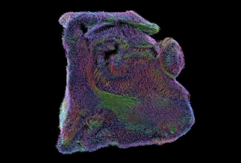
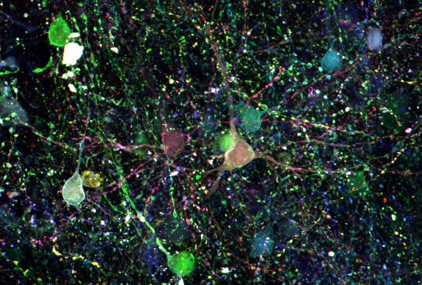
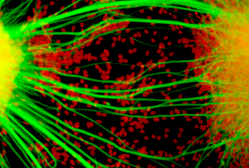
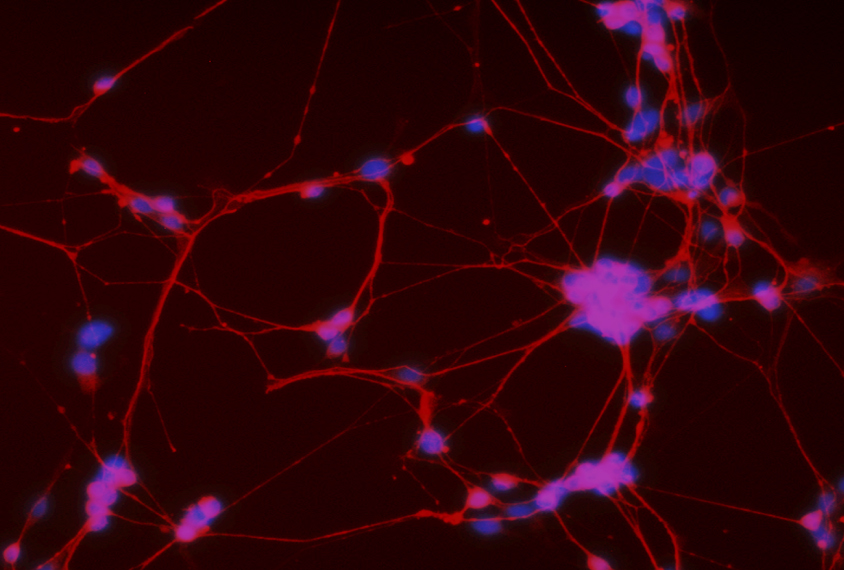
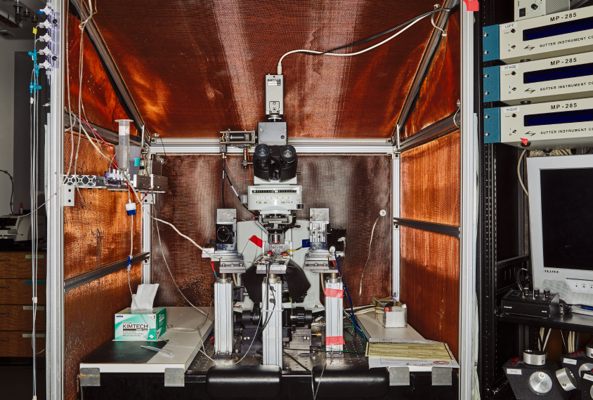
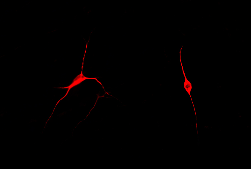
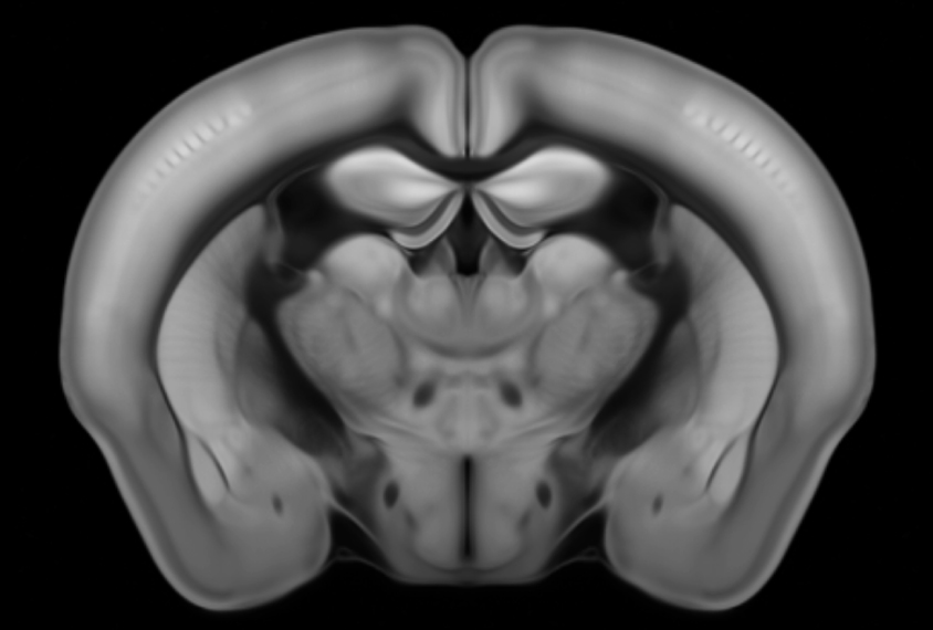
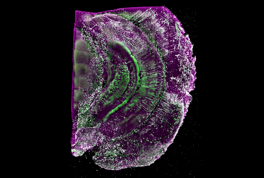
We asked autism researchers to enter the Spectrum science image contest. From sensational stem-cell snapshots to a ‘furry’ close-up of the hippocampus, these are the 10 top pics.


Fuzzy memories: A close-up of a human hippocampus, created by magnetic resonance imaging, shows the short-range connections critical for forming new memories.
Brainbow brilliance: Individual cells in the human brain reflect one of more than 100 hues after researchers treat the tissue with a combination of four dyes — a technique called ‘Brainbow.’
Creating connections: Two clusters of immature neurons (nuclei, red) derived from the cells of a child with autism form a bridge of fibers between them.
Magical mind: Data from magnetic resonance images of multiple human brains blend to form a colorful depiction of the bundles of nerve fibers (right) that transmit information in the brain.
In the pink: After seven days in culture, neural stem cells (pink) derived from a person with autism express a protein called beta-III tubulin (red) present in immature neurons.
Decoding device: This cell-recording machine captures the electrical activity of specific cells in slices of brain tissue that have been activated by light.
Simple cells: Neurons derived from people with autism have few branches (right). Exposing them to support cells called astrocytes (black) from controls leads to a more typical shape (left).
Rodent reveal: A 3-D model of a mouse brain shows more than 500 brain regions in great detail.
Painted cells: Three separate stains (green, red, blue) highlight precursors of human neurons grown in culture.
Rhapsody in green: Neurons infected with the rabies virus glow green in the hippocampus (memory hub) of the mouse brain.
0Comments
By joining the discussion, you agree to our privacy policy.