THIS ARTICLE IS MORE THAN FIVE YEARS OLD
This article is more than five years old. Autism research — and science in general — is constantly evolving, so older articles may contain information or theories that have been reevaluated since their original publication date.
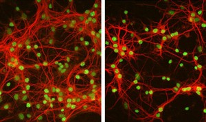
Diminished dendrites: Hippocampal neurons grown on a bed of astrocytes that lack MeCP2 have fewer and shorter dendrites (right), compared with those grown on normal astrocytes (left).
MeCP2, the gene that causes the autism-related Rett syndrome, is expressed not just in neurons but in glia ― cells that support neurons and help process information ― in the brain, according to a study published online in the March issue of Nature Neuroscience1.
What’s more, it’s MeCP2’s loss from these glia, and not from neurons, that appears to be important in causing the disorder. Glia make up roughly 90 percent of cells in the nervous system, and are thought to be crucial for the survival and maturation of neurons.
Rett syndrome is a rare neuro-developmental disorder that affects girls within the first 18 months of life. Beginning with delayed growth and speech, the disorder progresses and, as a result of severe impairments in motor function, often leaves girls wheelchair-bound by their early 20s.
A decade ago, Huda Zoghbi and her team first linked mutations in MeCP2 to the disorder2. Mutations in the same gene, which has been found to activate and repress thousands of other genes, are also found in people with learning disabilities, mental retardation and autism.
Several studies of Rett syndrome had reported MeCP2’s presence in neurons, but not in glia3. But a few years ago, some researchers began looking closer at the gene’s expression in glia because it is present in many other non-neuronal cell types ― such as fibroblasts and muscle cells ― outside the brain, says Nurit Ballas.
Ballas, who conducted the work as a research associate in the lab of Gail Mandel, is now associate professor of chemistry and cell biology at Stony Brook University in New York.
“I thought, ‘Why would I not see [MeCP2] in glia?'” Ballas recalls. She decided to use a sensitive antibody and a new way of staining for the protein that could enhance the signal.
Diverse locations:
She and her colleagues found that MeCP2 is present in all types of healthy mouse glia, including astrocytes ― star-shaped cells that help regulate a neuron’s chemical environment ― and oligodendrocytes, which form a protective sheath over neurons. In contrast, the protein is not detectable in the glia of mouse models of Rett syndrome.
Neurons cultured on a bed of astrocytes taken from Rett mice also have fewer and shorter dendrites ― the long fibers of neurons that receive signals ― compared with neurons cultured on healthy astrocytes.
Conversely, adding healthy astrocytes to neurons that lack MeCP2 restores the growth of dendrites in those neurons.
“To me the biggest surprise came from the fact that an astrocyte that doesn’t have this protein is compromised in its capacity to support neurons,” says Zoghbi, a Howard Hughes Medical Institute Investigator at the Baylor College of Medicine in Houston.
Even the astrocyte-conditioned medium ― a nutrient broth secreted by astrocytes that can normally support neuron growth in culture ― from the Rett mice stunts proper development of neurons.
The researchers are trying to selectively eliminate MeCP2 from mouse astrocytes and other glia in order to determine the relative roles of these cells in triggering Rett syndrome.
“We also want to know what the factors are in the media that astrocytes are secreting, and whether that plays a role in Rett,” Mandel says.
Because MeCP2 normally controls the expression of many genes, the lack of a functional MeCP2 in these astrocytes could stunt the neurons’ growth by somehow altering the chemical milieu in a neuron’s environment, the authors note.
“If it is true that the astrocytes are secreting a toxic inhibitory factor,” Mandel adds, “then that’s a point of potential pharmaceutical intervention.”
By joining the discussion, you agree to our privacy policy.