A multicolor labeling method stains neurons brightly enough to reveal the thin connections between individual cells1. Rendering the tissue transparent enables researchers to view the wiring of a mouse brain in three dimensions.
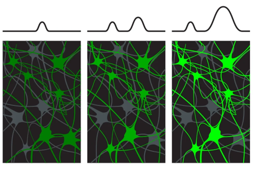
In the commonly used Brainbow approach, scientists use a virus to genetically modify neurons so that they produce different combinations of three fluorescent proteins — red, blue and green. Researchers broaden the palette by tweaking the number of gene copies for each color.
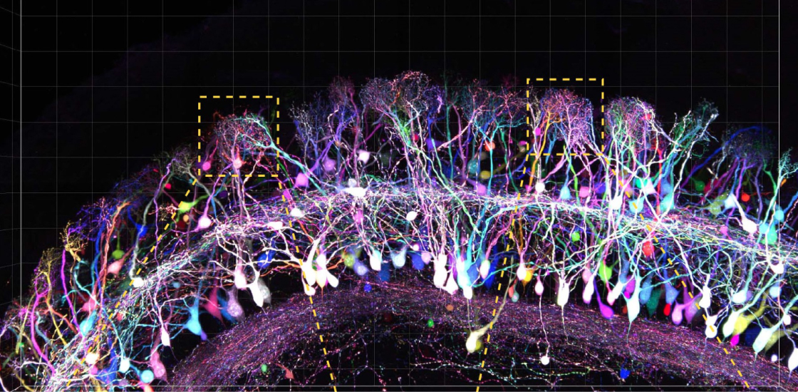
But the technique doesn’t stain neurons vividly enough to show their delicate projections — axons and dendrites. These projections may be altered in autism.
Hue variety: Staining each cell in a mouse brain region with a unique color makes neuronal connections easy to trace.
To boost the brightness, researchers added a strip of DNA that dials up gene expression so that each gene produces many more fluorescent proteins.
The researchers combined this ‘bright labeling’ with a technique they developed in 2016 that renders cells transparent by extracting fat molecules. They then traced neuronal projections as long as six millimeters from a mouse’s olfactory bulb, an interior brain structure that processes odors, to the cerebral cortex on the brain’s surface.
The researchers created videos from the images showing brilliant neuron cell bodies surrounded by tangles of fibers extending out in three dimensions.
The method is not perfect. Occasionally, a section of axon or dendrite does not produce the colorful proteins, creating a gap in the image. The researchers are working out ways to prepare their samples to eliminate these gaps.
Delicate detail: Minute neuronal extensions appear after brightly staining cells in the outer layer of a mouse brain.