A new microscopy technique offers the most detailed look to date at individual proteins present in small pieces of brain tissue, according to a new study.
Researchers presented the unpublished results virtually today at the 2021 Society for Neuroscience Global Connectome. (Links to abstracts may work only for registered conference attendees.)
Imaging the contents of synapses, the small and crowded junctions between neurons, has long been stymied by the limited capabilities of existing microscopes: Standard light microscopes are restricted in their resolution; more powerful microscopes, such as electron microscopes, cannot capture the colorful tags used to identify proteins in many studies.
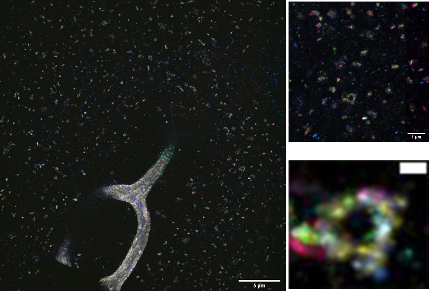
To get around these barriers, the researchers developed a new approach that enables them to repeatedly label a single tissue sample for multiple proteins. They combined this tactic with expansion microscopy, which improves image resolution by increasing the volume of the tissue. Together, the techniques provide a detailed view of the proteins present at synapses.
“With data like this, we can start to look at higher-dimensional relationships between proteins,” and potentially identify new subtypes of synapses, says Margaret Schroeder, a graduate student in the labs of Guoping Feng and Ed Boyden at the Massachusetts Institute of Technology, who presented the work.
The method is also compatible with Cre recombinase, Schroeder and her colleagues found. This enzyme can be used to test the effects of mutations in transgenic mice, including some mouse models of autism.
Label, rinse, repeat:
In the new work, Schroeder and her colleagues first used expansion microscopy on mouse brain tissue. Expansion microscopy, which the group initially developed in 2015, uses a water-absorbing gel to stretch a piece of tissue without damaging the structure of its cells. Bloating the tissue creates more space between molecules, increasing the resolution of the image by up to 20 times.
The researchers then stained the expanded tissue by using antibodies that bind to particular proteins. After imaging the tissue, they stripped it of those antibodies, using detergent and heat, and used new antibodies to stain the tissue again for different proteins.
Schroeder and her colleagues demonstrated that they could repeat this process at least four times, applying additional rounds of staining while maintaining the intensity of the image. As a result, they found they could image tens of proteins in the same field of view; current methods allow for imaging of only three to four proteins at a time.
Because they are capturing more data about synapses, the researchers plan to explore how proteins cluster in these regions. They say those results may provide new insight into synapse diversity and function.
Read more reports from the 2021 Society for Neuroscience Global Connectome.