THIS ARTICLE IS MORE THAN FIVE YEARS OLD
This article is more than five years old. Autism research — and science in general — is constantly evolving, so older articles may contain information or theories that have been reevaluated since their original publication date.
A new ultrasound device provides high-resolution movies of blood flow through the brains of infants1. Researchers could use it to identify and treat seizures, which occur in about one-third of children with autism.
Scientists typically measure brain blood flow using functional magnetic resonance imaging (fMRI). These scans can reveal which parts of the brain are affected by seizures. But the machines are large and expensive. They are also noisy and require participants to remain still, making them impractical for scanning infants.
To address these hurdles, researchers developed a portable ultrasound device that uses high-frequency sound waves to visualize brain activity. The device snaps more than 10,000 frames per second, compared with the 50 frames per second offered by traditional ultrasound scanners. This makes the device sensitive to changes in the flow of blood through small brain vessels.
The ultrasound probe weighs just 40 grams and rests on a flexible mount atop a baby’s head. Researchers position the probe to direct ultrasound waves through the fontanelle, the gap in an infant’s skull where the bones have not yet fused. Researchers described the device 11 October in Science Translational Medicine.
Spotting seizures:
The researchers used their device to measure blood flow in the brains of six infants, all about 10 days old. They also fitted the infants with eight electrodes for electroencephalography (EEG) to help assess the new device. EEG can diagnose seizure activity but cannot spot the location of seizures.
The new device was able to distinguish between quiet and active sleep states — which are well-defined stages associated with distinct EEG patterns — by recording changes in the volume of blood in the brain.
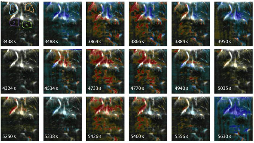
The researchers then used the device in combination with EEG in two newborns diagnosed with seizures. For both infants, the combined recordings showed how abnormal patterns of neuron firing spread to different parts of the brain.
The researchers were able to identify the exact location in the brain where the seizure activity started. The ultrasound images are of higher resolution than those captured with fMRI and infrared light waves, the researchers say.
By joining the discussion, you agree to our privacy policy.