THIS ARTICLE IS MORE THAN FIVE YEARS OLD
This article is more than five years old. Autism research — and science in general — is constantly evolving, so older articles may contain information or theories that have been reevaluated since their original publication date.
Using a sugar alcohol found in fruit, researchers have concocted a new chemical cocktail for making brains transparent.
In the past several years, scientists have devised various ways of making the brain transparent in order to explore its intricate circuitry. Many of these techniques end up harming the cells they are meant to examine, however.
A new method, described 14 September in Nature Neuroscience, offers a relatively benign way of rendering the brain see-through1.
Brains are opaque in large part because they are full of fats, which form cell membranes. In existing methods for preparing brains for imaging, researchers use high concentrations of harsh detergents or organic solvents to dissolve the fats.
Methods that use less harsh chemicals have other drawbacks. For instance, an early method called called ScaleA2 flushes fats out of the brain using a combination of urea, glycerol and a small amount of detergent. But urea causes the brain to swell and distort, and renders the brain only partially transparent.
In the new study, the researchers modified ScaleA2 by replacing glycerol with sorbitol — a sugar alcohol that makes tissues shrink. The resulting mixture of sorbitol and urea, called ScaleS, maintains the brain at its original size while making it transparent.
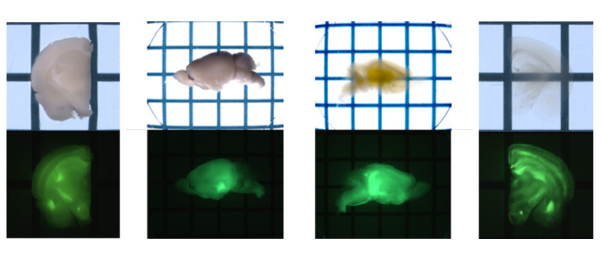
The researchers pitted the new formula against three older techniques in mice engineered to express fluorescent proteins in neurons. The older methods make neurons glow faintly because the harsh chemicals damage the cells. The researchers also tested a gentle fourth method and found that it preserves fluorescence but leaves brains fairly opaque.
ScaleS, by contrast, makes the brain transparent, with brightly glowing neurons. Electron microscopy revealed that unlike the older methods, ScaleS leaves cell membranes intact.
ScaleS may allow researchers to trace synapses, the connections between neurons, in three dimensions. Scientists could apply the technique to mouse models of autism or brain tissue from people with the disorder.
By joining the discussion, you agree to our privacy policy.