A metric called a polyneuro risk score can help researchers figure out relationships between brain activity and behavior, according to a new study.
Scientists could use the approach to characterize subtypes of autism and other neurodevelopmental conditions, although more research is needed, the study investigators say.
For decades, researchers have used functional MRI (fMRI) brain scans to try to link complex behaviors to variations in functional connectivity patterns. But such studies typically have insufficient sample size and statistical power to yield replicable results, says Oscar Miranda-Dominguez, assistant professor of clinical behavioral neuroscience at the University of Minnesota in Minneapolis and senior investigator of the new work. Because of publishing biases, negative associations between behavior and brain activity often go unpublished, he adds.
“We can’t estimate the real effect of an association just by doing a meta-analysis, because the information is missing,” he says. “Something is wrong with our current approach of studying brain-behavior associations.”
The issue is not unique to neuroscientists; geneticists faced a similar obstacle in their attempts to estimate disease risk based on variations at a single locus. Their solution was to calculate a polygenic risk score, which combines the cumulative small effects of multiple variants across the genome.
A similar approach could prove useful for studying the brain, a 2021 study found. The combined effect of small differences in brain activity across the cortex was a better predictor of a person’s performance on a behavioral task than was any single change in localized brain regions.
In the new paper, the researchers adopted a similar methodology but used a different set of parameters and mathematical formulae, explains Miranda-Dominguez. And unlike previous studies, their code is available online for others to use.
“It’s a data-driven approach that allows us to see what the brain is telling us, rather than putting our preconceived notions onto the data,” says lead study investigator Nora Byington, neuroimaging specialist at the University of Minnesota.
B
Using 3,339 of the scans, the team determined how strongly each brain connection relates to the participants’ general ability on a battery of cognitive tests. They then applied the calculated weights for all of the connections to the remaining scans to calculate a polyneuro risk score.
The top 10 percent of connections explained 16 percent of variation in participants’ general ability on the cognitive tests, far more than when the researchers randomly picked the same number of connections for their calculation. They obtained similar results when they reversed the order in which they analyzed the scans — using the second set to calculate weights and the first set to compute risk scores.
They saw less reproducibility when they applied the method to two other measures of cognitive performance — executive function and learning and memory — possibly because the identified connections have smaller effects on these skills, the researchers say. A larger sample size might be needed to study the relationship of these skills to brain activity, Byington says.
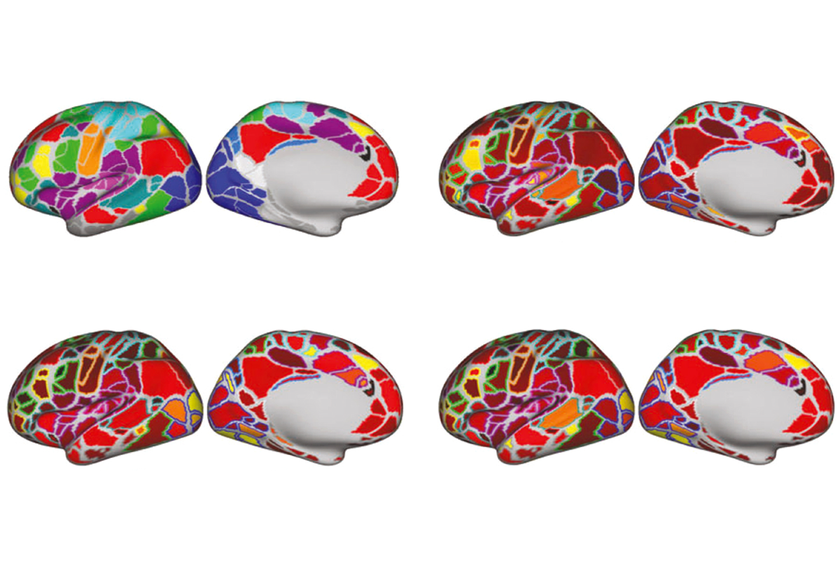
For all three cognitive measures, crosstalk between distal brain regions appeared to have a larger influence than local connections did.
The results were published online in March in Developmental Cognitive Neuroscience.
T
The framework could also help clinicians predict autism traits without the need for exhaustive testing, Miranda-Dominguez says. “You could have a number that is close to what you’d get from a trained professional. This is what we see for the future.”
But the team needs more data to test this idea. “We may not necessarily be powered to pick up on some clinical populations. This is something that we are working on,” Byington says.
“The jury’s still out on how it translates to psychiatric and neurological conditions,” says Scott Marek, assistant professor of radiology, also at Washington University. “We don’t have very large samples among these populations, and generating them will require multi-lab collaborations,” says Marek, who was not associated with the research.
Amassing enough brain images will require a “paradigm shift” toward collaborative work over individual research pursuits, Dosenbach says. A united effort, combined with the decreasing costs of MRI and improvement in motion correction techniques, could produce a mass of data, he says. “If the societal interest is there, you can collect thousands of datasets.”