THIS ARTICLE IS MORE THAN FIVE YEARS OLD
This article is more than five years old. Autism research — and science in general — is constantly evolving, so older articles may contain information or theories that have been reevaluated since their original publication date.
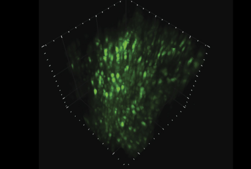
A new method allows researchers to watch thousands of neurons firing in concert in the brains of living mice1. The method provides a deeper, broader view of brain activity than traditional techniques, revealing interactions of neurons in networks.
The approach could allow scientists to learn whether neural networks are altered in mouse models of autism.
Researchers can track brain activity in mice by monitoring molecules that glow when neurons fire. They engineer mice to produce the molecules, which they excite with lasers. They then detect the glow with a specialized microscope mounted on a frame over a tiny window in the mouse’s skull.
Researchers have modified the approach to produce detailed images of individual neurons. But visualizing large numbers of neurons in three dimensions has been difficult: The glow from cells at different depths in the brain can overlap and obscure the overall picture.
In the new study, published 31 October in Nature Methods, researchers synchronized pulses of laser light to the time it takes to detect the resulting glow. This approach reduces the amount of fluorescence produced, speeding data collection and making possible the scanning of large areas. The scientists also aim the light toward the bulky cell bodies of neurons rather than their delicate branches, which further reduces out-of-focus glow.
The researchers designed and built a custom laser capable of providing the short pulses necessary for the method to work.
These innovations allowed the researchers to simultaneously see the activity of up to 4,000 neurons in 500 cubic microns of brain tissue — more than double the volume captured by existing methods.
The researchers tested the setup in mice as the animals ran on a rotating disk and reacted to puffs of air on their faces. The animals also wore a device that immobilized their heads. As the mice ran, the microscope captured firing from thousands of neurons across multiple layers of the posterior parietal cortex, which is involved in perception of the body in space. It revealed similar activity in the hippocampus, which encodes experiences for storage in memory.
By joining the discussion, you agree to our privacy policy.