THIS ARTICLE IS MORE THAN FIVE YEARS OLD
This article is more than five years old. Autism research — and science in general — is constantly evolving, so older articles may contain information or theories that have been reevaluated since their original publication date.
A new multi-step brain imaging method called ClearMap creates a snapshot of neuronal activity across the entire brain of an adult mouse1.
The tool, described 25 May in Cell, could help researchers understand how symphonies of neurons firing during social interactions differ between mouse models of autism and typical mice.
Scientists have used various methods, such as functional magnetic resonance imaging, to record synchronized activity in brain regions. But these techniques cannot identify which patterns of activity correspond with particular behaviors.
In the new method, a specialized microscope shines light on various depths of the mouse brain to produce a three-dimensional (3D) rendering of active neurons. New software then superimposes this image onto a publicly available atlas of brain structures. Scientists can analyze this combination to pinpoint the brain regions that fire in synchrony and match that activity to what the animal was doing just before they examined its brain.
Visual slices:
Researchers constructed this multi-step recipe from old and new techniques. First, they provoke brain activity, either with a drug or by engaging mice in a behavioral task. They then euthanize the animals and examine their brains.
Using a method called iDISCO+ , the researchers marinate the mouse brains in a chemical that renders them transparent. They label neurons that were firing while the mouse was alive using fluorescent antibodies that bind to a protein that a cell makes 30 minutes after it becomes active.
Next, a light-sheet microscope shines a plane of light through the brain to different depths and detects the glow of the recently active neurons in each visual slice. Software combines the scans into a single 3D image.
From there, a novel computer program called ClearMap that the researchers built overlays the 3D image onto the Allen Mouse Brain Atlas, pinpoints the location of activated neurons and measures the fluorescence in each region.
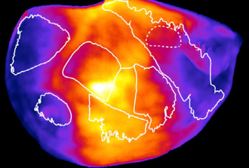
The scientists tested the technique in adult mice with shaved whiskers that had been exploring an unfamiliar area. The tool revealed low activity in brain regions that receive information from whiskers, and unusually strong activation among neurons that pick up sensory signals from the nose and mouth.
In another experiment, the platform displayed interactions among three brain regions in mouse mothers that had been moving their pups to a nest: a part of the hypothalamus involved in parenting behavior; the thalamus, a sensory relay station; and the amygdala, an emotion hub.
The iDISCO+ and ClearMap tools are freely available online.
By joining the discussion, you agree to our privacy policy.