A new microscope creates images of a mouse embryo as it grows from a ball of about 100 cells to a structure with a primitive heart and brain1.
From these images, researchers were able to compile an atlas showing where thousands of cells begin and end up in this process.
The resource could help scientists understand how and where cells migrate during development, and how that process is altered in conditions such as autism.
Imaging living embryos is a major technical challenge. Exposure to light from a microscope can alter development; the microscope also needs to continually refocus to obtain clear images of an embryo’s rapid growth and structural changes.
The new microscope includes a culture system that nurtures a 6-day-old mouse embryo. The embryo floats in a broth of nutrients in a small chamber at the center of the microscope. As the embryo drifts in the media, growing and changing shape, custom hardware and software move the microscope’s lenses and areas of focus to keep the embryo in view.
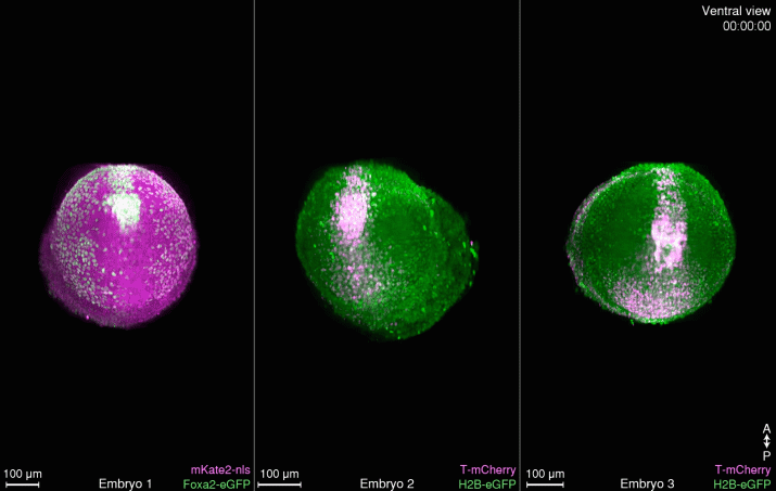
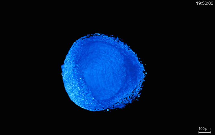
Center stage: A mouse embryo’s ‘notochord plate,’ which will morph into part of the spinal cord, elongates between days 6 and 8 of development.
K. McDole et al. / Cell 2018
Other algorithms adjust the illumination to adapt to changing tissue depth. And time-lapse techniques capture the changes while minimizing the amount of light needed for a picture.
Using this system, the researchers captured images of mouse embryos for 48 hours as the embryos grew to about 250-fold their original size. The researchers created software to extract position information for thousands of individual cells from the 10 terabytes of images. With those data, they built an interactive 3-D atlas showing the migration path of each cell.
The atlas reveals structures that morphed into the heart, brain and other organs at single-cell resolution.
It also shows dynamic developmental processes such as the zipper-like closure of the neural tube, a hollow structure that becomes the brain and spinal cord. Researchers described the resource in October in Cell.
The microscope is housed at the Advanced Imaging Center at the Janelia Research Campus in Ashburn, Virginia, where scientists can use it for free. A team of experts is available to help users prepare samples, create images and perform data analysis.