THIS ARTICLE IS MORE THAN FIVE YEARS OLD
This article is more than five years old. Autism research — and science in general — is constantly evolving, so older articles may contain information or theories that have been reevaluated since their original publication date.
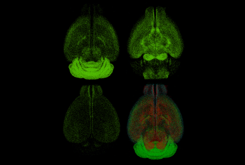
A new collection of maps reveals the distribution of inhibitory neurons across the entire mouse brain1. Inhibitory neurons keep brain activity in check, and this delicate balance may be altered in autism.
Scientists can use fluorescent proteins to label specific types of cells and visualize them through a microscope. But a microscope’s field of view can capture only a small portion of the mouse brain at once. This makes the mapping of cell types across the brain a daunting task.
In the new study, researchers streamlined the process by combining automated tissue-imaging techniques with custom algorithms. They sliced the brains of mice that express fluorescent proteins in seven types of inhibitory neurons. They then used an automated microscope to illuminate the fluorescent proteins and capture multiple images of each slice.
The team created computer algorithms to analyze the images and count the glowing cells. They then mapped the cells onto a 3-D digital rendering of a mouse brain that is based on data from the Allen Mouse Brain Atlas.
The approach yielded the numbers of inhibitory neurons within more than 800 brain regions, according to results published 5 October in Cell.
Marking territory:
The numbers and types of inhibitory neurons vary by brain region. One type of inhibitory neuron fires rapidly to suppress its target neurons; another fires at a rate that depends on feedback from target neurons. The researchers found that the first type dominates motor regions of the brain’s surface, the cerebral cortex. The second is abundant in the association cortex, an area involved in processing sensory information.
The researchers used their maps to build computer models of mouse brain circuits and to run simulations of neuronal activity. The simulations revealed activity patterns that correspond to the ratios of different inhibitory neuron types.
The team also found that female mice have more inhibitory neurons than males do in nine brain areas. These areas are involved in sensing smells, processing pheromones and social behavior.
The brain maps and the data they include are freely available online. The software is set up to allow scientists to add datasets for other cell types, such as excitatory neurons, and for mouse models of conditions such as autism.
By joining the discussion, you agree to our privacy policy.