Landmark atlases flag 5,000 cell types across mouse brain

The work identifies new varieties and may help researchers develop tools to genetically target specific classes of cells.

Six cross-sections of mouse brains.
Images courtesy of Cindy van Velthoven, Michael Kunst and Delissa McMillen at the Allen Institute

Two new atlases catalog the location and type of all cells across the adult mouse brain — including many that have never before been identified, according to two new unpublished studies.

The resources represent “a culmination of landmark efforts” to try to establish a census of cell types, says Tomasz Nowakowksi, associate professor of neurological surgery at the University of California, San Francisco, who was not involved in the work. “It’s presumably something that has been a dream since the days of Santiago Ramon y Cajal.”

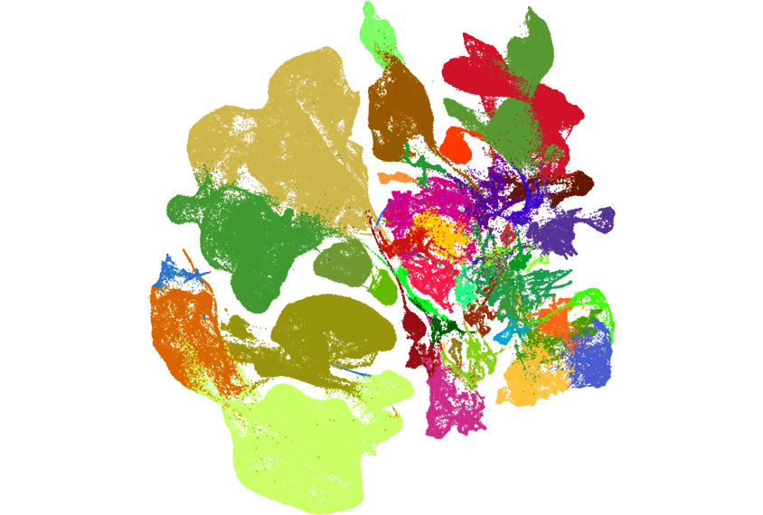
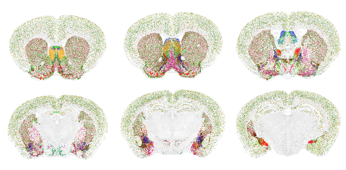
In an effort spanning more than 10 years, and using different approaches, two teams of researchers each identified about 5,000 distinct clusters that represent different kinds of cells.

The “tremendous diversity” they observed was surprising, says Hongkui Zeng, executive vice president and director of the Allen Institute for Brain Science in Seattle, Washington, who led the work on one of the new atlases. Some of the clusters map to known cell types, Zeng says, but others “we never heard about before.”

That new information will help guide future studies, Nowakowski says. “Imagine that you’re trying to sail across the ocean, and you don’t really have a map,” he says. “That’s exactly what’s been happening with neuroscience for the past 100 years.”

T

o develop that map, Zeng and her colleagues performed single-cell RNA sequencing on tissue from adult mouse brains. They identified 5,200 distinct clusters of cells from the RNAseq data, and then picked out the pattern of gene expression — a “molecular handle,” as Zeng calls it — that best distinguished one cluster from another.

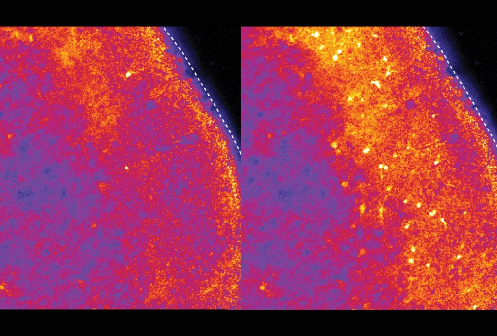
The team also located the RNA molecules within the same slices of brain tissue using a method called multiplexed error-robust fluorescence in situ hybridization (MERFISH), which associates each RNA with a barcode linked to a particular spot. They then flagged which cell-type clusters appeared at a given spatial location using the previously obtained molecular handles.

Research image of cell types in the mouse brain.
Type set: Groups of cells in the mouse brain can be identified by their specific patterns of RNA expression.

Specific cell types tended to cluster in particular brain regions, the team found. Those cell types frequently lined up with ones neuroscientists have already been studying, such as as a class of cells in the hypothalamus known to be important for innate behaviors, Zeng says. And most cell types tend to express a single neurotransmitter, based on the neurotransmitter genes associated with each cluster.

“It just made us believe that this approach is really working,” Zeng says.

The second team, led by Evan Macosko, associate professor of psychiatry at Harvard Medical School and institute member at the Broad Institute in Cambridge, Massachusetts, used slightly different methods to construct their maps. They sequenced the RNA data from each cell’s nucleus, rather than from the entire cell, and then mapped the corresponding spatial information using Slide-seq, a method that transfers a cell’s RNA onto barcoded beads to locate the RNA in space.

Despite their differences, the two groups came to similar conclusions, Macosko says. And because the atlases took a different focus, with Zeng’s work including more data from the brain’s cerebral hemispheres and Macosko’s including more from other regions, such as the brainstem, combining them could also prove useful, he says.

Both studies stem from the National Institute of Health’s Brain Initiative Cell Census Network and were posted to bioRxiv last month.

I

n addition to revealing the large diversity of cell types within the brain, the new work shows that that diversity is not uniformly distributed, Macosko says. “There’s an enormous amount of specialization in the brainstem,” he says, as well as in the hypothalamus, midbrain, medulla and pons.

That discovery may encourage more researchers to investigate these understudied brain regions, Macosko adds, and the new atlases could provide them the toolkit to do it.

One current challenge is that there is no way to genetically target specific cell types in, for example, a mouse’s brainstem. But the new atlas from Macosko and his colleagues identifies “the minimum number of gene combinations that are needed to distinguish every cell type within a brain area and across the whole brain,” he says — essentially a recipe book for how to genetically target any cell.

For that reason, the new resources open the door to new studies of cell function, and even to the development of therapeutics targeting specific cell types, Nowakoswki says.

The next frontier is developing the same type of atlas for the human brain, he says — something that both Zeng and Macosko say they plan to take on next.

“Humans have many more areas, and only some of the cell types are conserved across areas, others are not,” Nowakowski says. The question is, then, “how much complexity will we discover when we look at the human brain?”