M. Renner et al. / The EMBO Journal
THIS ARTICLE IS MORE THAN FIVE YEARS OLD
This article is more than five years old. Autism research — and science in general — is constantly evolving, so older articles may contain information or theories that have been reevaluated since their original publication date.
A new method for examining lab-grown ‘mini-brains’ reveals structures like those in the human brain1.
Mini-brains, also known as cerebral organoids, can provide clues to brain development. To build them, scientists coax clusters of stem cells into becoming neurons and other brain cells. They can even start with skin cells from a person with autism to see how the person’s genes influence the mini-brain’s structure. But researchers debate how closely mini-brains resemble human brains.
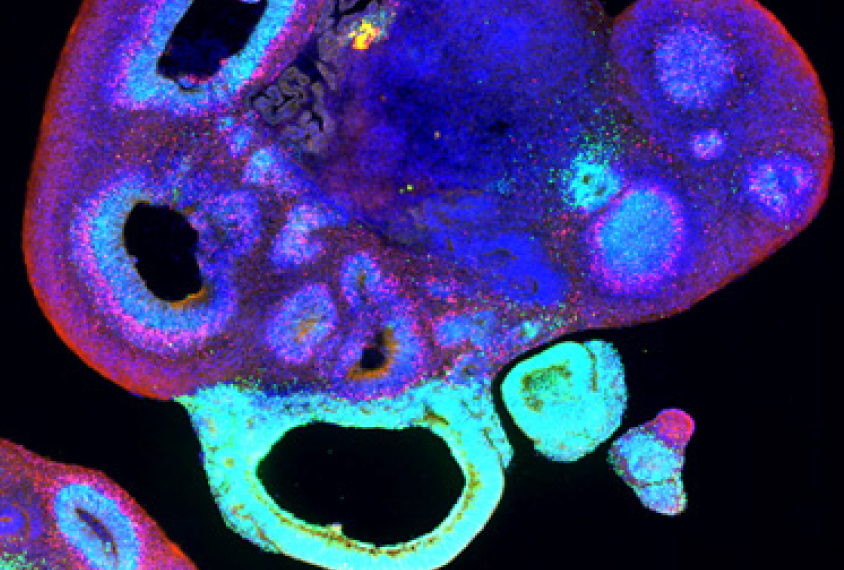
In the new study, published 10 March in The EMBO Journal, researchers probed the cellular and regional structure of 104 mini-brains grown from human embryonic stem cells. They first froze the mini-brains and cut them into ultra-thin sections, which they mounted onto glass slides. They then labeled the sections with different combinations of colored fluorescent tags that are specific to certain cell types, and imaged the sections using an automated scanner.
The tags revealed a mixture of cells, including mature neurons and star-shaped support cells called astrocytes. The mini-brains are irregular blobs with small inner chambers, but the researchers found that they contain complex tissues.
Organizing centers:
A region within each mini-brain resembles the human forebrain, which governs complex cognitive tasks such as integrating sensory information. This region often develops as a folded, ribbon-like structure near the outside of the organoid. It contains layers of cells like those seen in the human cortex.
The researchers used a chemical cocktail to render some of the mini-brains transparent. This revealed bridges of tissue that connect different parts of the forebrain-like region.
The researchers also examined mini-brains at various time points from 33 to 160 days old, when their cells are fully mature. The cells matured into neurons and other brain cells at a speed and in a sequence similar to those in the developing human brain.
Some mini-brains formed patches of cells that secrete chemical cues that spur the development of certain cell types or delineate regions. These patches are similar to so-called ‘organizing centers’ in the developing human brain.
The method revealed significant variability in the size and location of the forebrain. This may arise from when and where the organizing centers form, or whether they form, the researchers say.
By joining the discussion, you agree to our privacy policy.