THIS ARTICLE IS MORE THAN FIVE YEARS OLD
This article is more than five years old. Autism research — and science in general — is constantly evolving, so older articles may contain information or theories that have been reevaluated since their original publication date.
A new method offers an efficient way to examine gene expression in individual cells from postmortem brain tissue1.
Scientists typically measure gene expression by looking at messenger RNA (mRNA), the intermediary between DNA and protein. Traditional methods for doing this require large batches of cells, and yield only crude estimates of the expression in a single cell.
Researchers can also sequence mRNA in individual cells, but their approaches mainly work with fresh cells, not chemically preserved or frozen ones.
The new method combines two existing techniques: One pulls cell nuclei from preserved tissue, and the other sequences mRNA from tens of thousands of individual nuclei at once. Isolating nuclei allows researchers to forego the harsh chemicals used to tease RNA from tissue. This leaves more of the RNA intact and the sample less biased toward certain cell types.
The researchers tested the approach on postmortem brain tissue from mice and people; the human tissue had been frozen for up to 5 ½ years. They took samples from the hippocampus, which plays a role in memory; and the prefrontal cortex, which is involved in attention, judgment and decision-making.
Signature signs:
In the method, researchers grind up chunks of tissue, add enzymes to break up the cells, and sift the nuclei. They inject the nuclei, which are suspended in liquid, into a glass chip etched with channels.
This stream of nuclei meets another carrying plastic microbeads, and then merges with a trickle of oil. The physical properties of the system cause the oil to coat and encapsulate a single nucleus and microbead in a microscopic droplet.
The microbeads are studded with small strands of DNA. These strands stick to mRNAs in the nuclei, acting as barcodes that connect the mRNAs to the cell they came from. After breaking apart the droplets and pooling the samples, scientists can sequence the RNA transcripts and the attached barcodes. The process reveals which genes are active in each cell.
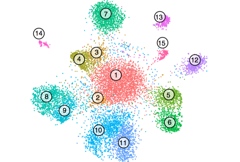
The researchers classified dozens of neuronal and other cell types in the hippocampus and prefrontal cortex according to their known gene expression signatures. They found subtle distinctions between types of mouse and human neurons, and even picked up rare cell types such as neural stem cells. They described the technique 28 August in Nature Methods.
Scientists could use the method to compare gene expression in specific cell types in postmortem brain tissue from people with autism.
By joining the discussion, you agree to our privacy policy.