THIS ARTICLE IS MORE THAN FIVE YEARS OLD
This article is more than five years old. Autism research — and science in general — is constantly evolving, so older articles may contain information or theories that have been reevaluated since their original publication date.
A modified version of an existing brain-imaging technique creates crisp, three-dimensional renderings of neurons communicating in living mice1. Researchers can use the technique to home in on delicate neuronal structures.
Scientists can track brain activity in living mice by engineering the animals to produce molecules that glow when neurons fire. This technique requires cutting a small window in a mouse’s skull and aiming lasers at the neurons to excite the molecules.
This method works well for visualizing active neurons in a single plane, but does not adjust the depth of the laser quickly enough to capture neuronal firing in three dimensions. What’s more, slight movement from breathing or blood flow through nearby vessels can blur the images. Fine structures such as the signal-receiving branches of neurons, called dendrites, are particularly challenging to scan.
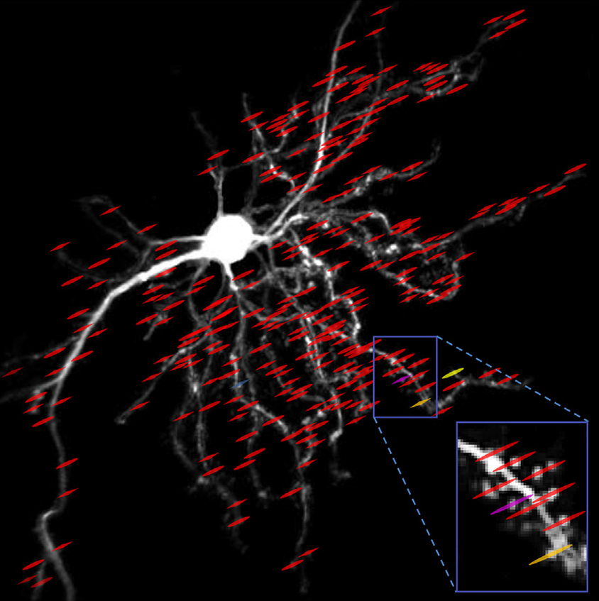
In the modified setup, described 20 October in Neuron, the properties of the laser light change rapidly to capture neuronal activity at different depths. The update also includes computer algorithms that correct for movement. Scientists can examine firing neurons at various scales, zooming in on part of a neuron or panning out to visualize groups of neurons.
The researchers tested the new method in mice as the animals responded to sounds or ran along a treadmill and navigated a virtual maze projected on a screen in front of them. The researchers recorded spikes of fluorescence in more than 150 dendritic spines of a single neuron. Separately, they captured activity across a network of more than 100 neurons at a higher resolution than has been possible before. These capabilities should provide new ways to study neuronal signaling and network connections, the researchers say.
Scientists can upgrade their microscopes to use the new method by refining electronic circuits and adjusting the mirrors that shape the laser beam, among other modifications.
By joining the discussion, you agree to our privacy policy.