THIS ARTICLE IS MORE THAN FIVE YEARS OLD
This article is more than five years old. Autism research — and science in general — is constantly evolving, so older articles may contain information or theories that have been reevaluated since their original publication date.
Researchers have mapped the migration patterns of neurons in the developing monkey brain and pinpointed when the neurons establish their identities. The unpublished results, presented yesterday at the 2015 Society for Neuroscience annual meeting in Chicago, suggest that neurons find their homes early in development but mature much later on.
“They continue to have gene expression changes throughout development,” says Jeremy Miller, scientist with the Allen Brain Institute in Seattle, Washington, who presented the work.
The results are the latest update to the Blueprint Non-Human Primate Atlas — a National Institutes of Health initiative to map brain gene expression in the rhesus macaque from 40 days after conception to 4 years of age.
Miller and his colleagues first looked at the layered structure of the brain’s outer rind, called the cortex, at six prenatal time points and four after birth. They found that many neurons in monkeys have finished the journey to their lifelong layer roughly 80 days after conception — the middle of the second trimester.
The researchers then isolated individual neurons from these layers using laser capture micro-dissection and measured their gene expression. They found that neurons express different genes prenatally than they do after birth. In fact, only 60 percent of genes that distinguish between neurons from different cortical layers in 1-year-old monkeys continue to do so in 4-year-old animals, according to their analysis.
“The further back you go in time, the less similar the gene expression pattern,” says Miller.
Delayed maturity:
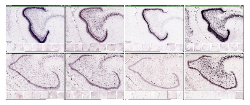
There is some evidence that neurons at the front of the brain develop earlier than those at the back. To explore this possibility, the researchers homed in on the anterior cingulate gyrus (ACG) — a C-shaped structure in the brain’s frontal lobe — and the primary visual cortex (V1), a patch on the back of the brain.
They found that tissue from the ACG expresses genetic markers for neurons and star-shaped support cells, called astrocytes, as early as 70 days after conception. By contrast, tissue from V1 expresses only cell-cycle markers at this time point, suggesting that neurogenesis is just getting started.
The ACG is involved in learning, memory and emotion, and there is some evidence that it is disrupted in children with autism. The new findings suggest that the brain structure develops during the first and second trimester, adding to mounting evidence that mid-fetal development is a critical period for autism risk.
To see how the findings from rhesus macaques jibe with those from people, the researchers compared their findings with data from the BrainSpan Atlas — a catalog of gene expression data from pre- and postnatal postmortem human brain tissue. They found that expression patterns for roughly 80 percent of genes were highly correlated between the two species.
The researchers are analyzing tissue from other brain regions implicated in autism, including the prefrontal cortex, the striatum, the amygdala and the hippocampus. The findings could help researchers identify developmental periods that underpin the disorder.
For more reports from the 2015 Society for Neuroscience annual meeting, please click here.
By joining the discussion, you agree to our privacy policy.