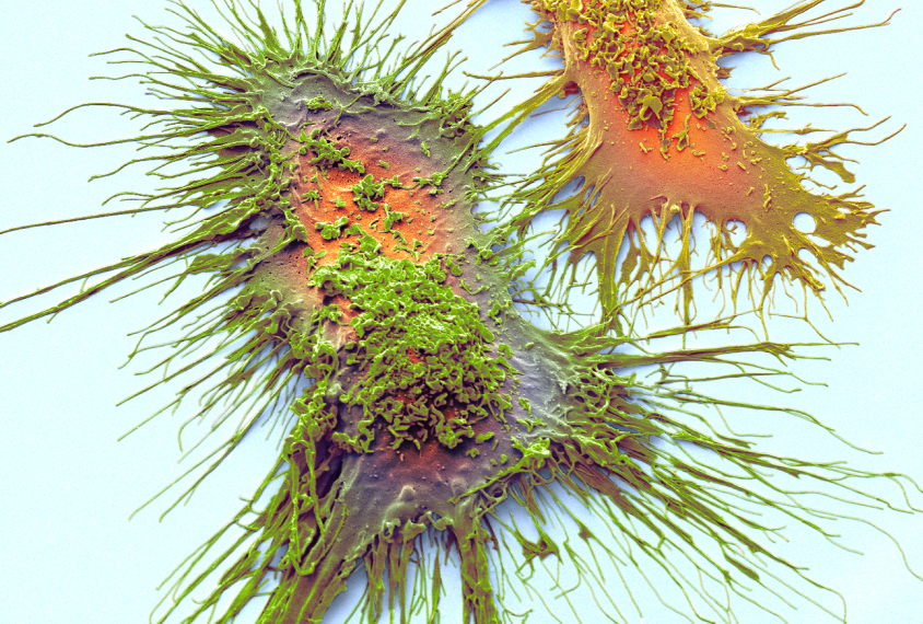
Steve Gschmeissner / Science Photo Library
THIS ARTICLE IS MORE THAN FIVE YEARS OLD
This article is more than five years old. Autism research — and science in general — is constantly evolving, so older articles may contain information or theories that have been reevaluated since their original publication date.
Immune cells in the brain called microglia play diverse roles in the mouse embryo, but assume a common function by adolescence. Researchers presented a map of the cells’ roles yesterday at a conference at the Broad Institute in Cambridge, Massachusetts.
The map provides a baseline against which scientists can compare the behavior of microglia in mouse models of autism, says Tim Hammond, postdoctoral researcher in Beth Stevens’ lab at the Broad Institute’s Stanley Center for Psychiatric Research. Hammond presented the findings.
Knowing the roles of microglia over time may help reveal whether the cells have a causal role in autism. In particular, microglia’s ability to prune neuronal junctions, or synapses, may be altered in the condition, which could shift the brain’s fundamental wiring.
Little is known about how microglia change during typical brain development. Hammond and his colleagues analyzed microglia in mouse brains at three stages: as embryos, a few days after birth and during mouse adolescence, which is around 30 days after birth.
They used a single-cell sequencing technique to analyze more than 75,000 individual microglia ‘transcriptomes,’ the array of genes expressed in the cells. By examining the transcriptomes at each stage, they inferred the cells’ roles at that point in time.
“What we find is there are at least nine different states that microglia can assume over the course of its development,” Hammond says.
Varied roles:
For instance, in the embryo and shortly after birth, the cells assume various forms that allow them to dispose of debris or become involved in immune signaling, cell division, cell movement or other functions.
“But as the brain matures, they become more and more like each other until they form one large cluster of microglia that are basically all the same,” Hammond says.
By the time the mouse is a month old, the researchers found, the cells are performing their essential action: patrolling for invaders. At this stage, the microglia are also likely snipping synapses, Hammond says, “but we don’t know for sure.”
The researchers plan to conduct single-cell sequencing of microglia in a mouse model of maternal infection, to determine how the cells respond in this scenario. They also plan to look for markers that could help identify microglia activity in people. These experiments could give clues about how microglia function in people with autism.
By joining the discussion, you agree to our privacy policy.