THIS ARTICLE IS MORE THAN FIVE YEARS OLD
This article is more than five years old. Autism research — and science in general — is constantly evolving, so older articles may contain information or theories that have been reevaluated since their original publication date.
Infants born prematurely show alterations in the structure and function of their brain circuits — findings that hint at why they are at increased risk for autism. Researchers presented the unpublished results, from two independent long-term studies, at the 2015 Society for Neuroscience annual meeting in Chicago.
One team also presented evidence that a low-tech intervention might mitigate the autism risk in these children.
Children who were born prematurely, meaning before 37 weeks gestational age, are at increased risk of autism, attention deficit hyperactivity disorder (ADHD) and other developmental problems. But the mechanism underlying this observation is unclear.
In the first study, the researchers scanned the brains of 76 infants who had been born at less than 30 weeks’ gestation around each baby’s due date. They also scanned 58 full-term infants within a few days after birth.
The researchers analyzed structural connections, called white matter tracts, in the infants’ brains. White matter contains nerve fibers that transmit signals between different brain areas. Many of these white matter tracts are less organized in preterm infants than in the full-term controls, the researchers found.
The team also assessed functional connectivity in seven brain networks — an indicator of how well disparate brain regions work together. They found that these networks are less coordinated in preterm infants than they are in controls, says study leader Cynthia Rogers, assistant professor of child psychiatry at Washington University in St. Louis, who presented the results yesterday at the conference.
In full-term infants, some pairs of networks are tightly related with each other in their activity, whereas others are uncoupled. Preterm infants show fewer of these positive and negative relationships between networks. “At this point it looks like they’re underdeveloped,” Rogers says.
The most pronounced changes are seen in the frontoparietal control network — which is involved in managing goal-directed actions — and the default mode network, a set of brain regions active when a person is awake but at rest. “These are some networks that show differences in children with ADHD and autism,” says Rogers.
Brain biomarker:
This idea that brain activity in premature infants is relatively unrefined is tantalizingly similar to a possible biomarker for autism risk identified in the second study.
In this study, researchers recruited the families of babies born at 24 to 36 weeks of gestational age. Of 150 babies, 72 received standard care in the neonatal intensive care unit and 78 received standard care plus Family Nurture Intervention, a program designed to strengthen emotional bonds between the premature baby and its family members, especially its mother.
This program has mothers of preterm infants place cloths that smell like their own skin in the baby’s incubator, and receive cloths that smell like their babies. They spend time speaking to the baby in emotional tones. Physical contact is also important: even for babies too fragile to hold in arms, mothers can place a finger against the baby’s palm to trigger the grasp reflex or gently but firmly press both hands around the baby’s torso, a calming touch.
The researchers have found that infants who receive this intervention have better cognitive and language skills at 18 months old than do those who receive standard care. They also have better scores on an autism-screening questionnaire called the Modified Checklist for Autism in Toddlers (M-CHAT).
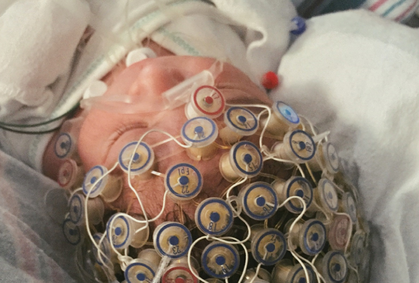
The team scanned the babies’ brains with electroencephalography (EEG), a noninvasive technique that measures electrical activity with a net of 128 electrodes worn on the scalp, when the babies were the equivalent of 37 to 44 weeks gestational age.
Coherent scores:
In both the intervention and standard care groups, an EEG measure known as coherence decreases as the babies get older. Coherence reflects similarities in the activity measured by any two electrodes in the EEG cap.
“Low coherence indicates greater independence and flexibility of function” of different brain regions, says study leader Martha Welch, director of the Nurture Science Program at Columbia University Medical Center in New York and chief architect of the intervention. “Early coherence in preterm infants decreases with maturation, indicating growing flexibility.”
Infants who receive the nurturing intervention have lower EEG coherence than do those who receive standard care1. “We see that finding already at term, only a few weeks into the intervention,” says Julia Chafkin, a research assistant in the Nurture Science Program, who presented the work on Sunday.
The researchers then analyzed the relationship between early EEG coherence and later scores on the M-CHAT as well as tests of cognition and language. They found that among infants who receive standard care, those with greater EEG coherence at term have poorer outcomes at 18 months. But among those who receive the intervention, there is no relationship between EEG coherence and later neurodevelopment. “We think that [the intervention] may mitigate the otherwise deterministic effects of early brain activity on long-term development,” Welch says.
Risk relationships:
The results suggest an unexpected role for coherence in signaling autism risk. And the intervention’s effect on M-CHAT scores is particularly surprising, says April Benasich, director of the Infancy Studies Laboratory at Rutgers University in Newark, New Jersey.
“You might expect improvement in cognitive measures, but not necessarily autism risk,” says Benasich, who was not involved with the work. EEG data from more frequencies would help to better interpret the link between coherence and M-CHAT scores, she adds. Without that fuller context, “it’s difficult to sort out what the pattern means in terms of what differs between these groups.”
Welch says her team is planning to explore other EEG measures. The researchers reported last year that their intervention disrupts a relationship between autism risk and two EEG biomarkers in children born prematurely. “We keep seeing things like this time and again with this group” of children, Chafkin says.
Rogers, who was not involved in this study, says the apparent effectiveness of the intervention is easy for her to accept. “I’m a wholehearted believer in the impact of caregiver interaction with infants to promote positive brain development,” she says.
Rogers and her colleagues are also tracking the children in their study to see how their early brain measures relate to their later development. For example, the team reported last month that in preterm infants, less organization in the left cingulum — a C-shaped bundle of white matter fibers deep in the brain — at the due date tracks with better scores on a measure of social and emotional behavior at age 22.
The children in both new studies have now reached 4 to 5 years of age and are returning for follow-up assessments. Children’s developmental strengths and weaknesses are often clearer at this age than they are in toddlerhood, Rogers points out. “Our five-year findings we think are going to be even more interesting.”
For more reports from the 2015 Society for Neuroscience annual meeting, please click here.
By joining the discussion, you agree to our privacy policy.