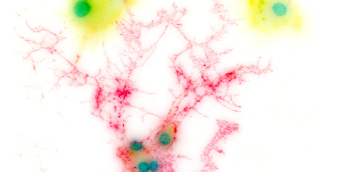
Riccardo Cassiani-Ingoni / Science Source
THIS ARTICLE IS MORE THAN FIVE YEARS OLD
This article is more than five years old. Autism research — and science in general — is constantly evolving, so older articles may contain information or theories that have been reevaluated since their original publication date.
Tiny pulses of blue light dampen the expression of inflammatory genes in microglia, the immune cells of the brain. The preliminary results, presented today at the 2015 Society for Neuroscience annual meeting in Chicago, highlight a possible side effect of optogenetics — a technique that uses blue light to activate specific subsets of neurons.
“One of the big selling points of optogenetics is that it’s so targeted,” says Kevin Cheng, a graduate student in Kevin Eliceiri’s lab at the University of Wisconsin-Madison, who presented the work. “But we’re showing that there are effects on other cells.”
Microglia are thought to play an important role in a number of neuropsychiatric conditions, including autism. The new findings raise the possibility that optogenetics experiments alter microglia’s effects on neurons — the very cells that researchers are trying to investigate.
“Whether or not there are other side effects in other cell types, we don’t know yet,” Cheng says.
Cheng and his colleagues cultured microglia from mouse brains and exposed them to lipopolysaccharide (LPS), a molecule that mimics an infection and spurs the microglia to action. They then flashed blue light on the cells at doses ranging from 1 to 100 millijoules per square centimeter — amounts similar to those used in optogenetics experiments.
Six hours later, the researchers sequenced RNA from the microglia to measure their gene expression. Microglia exposed to blue light show lower levels of a range of inflammatory genes, compared with untreated cells. These include TNF-alpha and COX-2, which encode powerful inflammatory molecules.
Light-treated, activated microglia also express increased levels of vascular endothelial growth factor (VEGF), which has protective effects on neurons.
Blue light has minimal effects on resting microglia that have not been exposed to LPS. The researchers suspect that the light triggers a cascade of signals in activated microglia that dampen the expression of NF-kappaB — a transcription factor that regulates other inflammatory genes.
Cheng and his colleagues are developing a method to measure blue light’s effects on microglia in living mice. They also plan to look at blue-light-induced gene expression changes in other brain cells, such as astrocytes, oligodendrocytes and even neurons.
For more reports from the 2015 Society for Neuroscience annual meeting, please click here.
By joining the discussion, you agree to our privacy policy.