Science Picture Co / Science Source
THIS ARTICLE IS MORE THAN FIVE YEARS OLD
This article is more than five years old. Autism research — and science in general — is constantly evolving, so older articles may contain information or theories that have been reevaluated since their original publication date.
Editor’s Note
This article was originally published 15 November 2017, based on preliminary findings presented at the Society for Neuroscience annual meeting. We have updated the article following publication of the study on 22 August 2018 in Neuron1. Updates appear below in brackets.
Researchers have charted billions of synapses in the mouse brain and sorted them by type, according to unpublished findings presented today at the 2017 Society for Neuroscience annual meeting in Washington, D.C.
They used a new technique that captures and categorizes these tiny gaps between neurons by labeling and sorting the proteins that line them. The method involves a new way to illuminate the proteins at synapses and a machine-learning method to analyze them. The resulting ‘synaptome’ is a comprehensive catalog of synapses in the mouse brain.
The technique could be used to map synapses in mouse models of conditions such as autism.
“We can apply this method to any disease model, in principle,” says Zhen Qiu, a postdoctoral researcher in Seth Grant’s lab at the University of Edinburgh in Scotland.
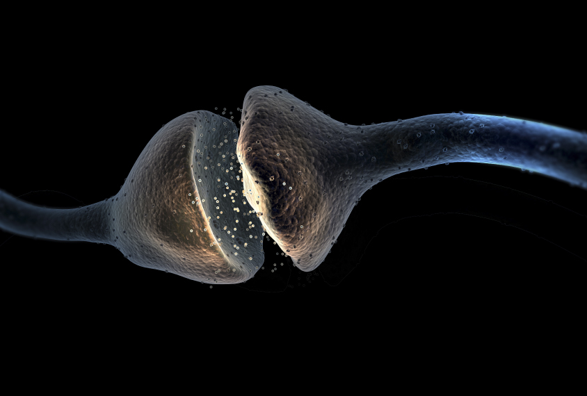
Synapses are junctions between neurons, and they mediate the transfer of information between the cells. Abnormalities in proteins at synapses have been implicated in autism and other neurodevelopmental disorders.
Some of these proteins are on the neuronal branch that sends the signal, and others are on the neuron that receives it. The researchers injected into the brains of mice florescent markers that attach to proteins at the receiving end. The markers reveal the number of parts, or subunits, that make up the protein by the intensity of the light emitted. And the pattern of fluorescence indicates each protein’s shape.
Sorting synapses:
The researchers used a spinning disk confocal microscope to capture the fluorescence. The result is a detailed picture of the location, size, shape and other features of these proteins.
The method is fast: Imaging an entire mouse brain took just half a day, Qiu says.
“That is feasible and practical for our study because we are doing hundreds of brains in our experiments,” he says.
The researchers wound up with terabytes of data. So they developed a machine-learning technique that automatically detects and categorizes the proteins based on the pattern of florescence. For instance, the software could determine how round or oblong the proteins are. It could decipher the proteins’ size and pinpoint the number of synapses within various brain regions.
The program then used the characteristics of proteins at each synapse to sort the junctions into categories. [The researchers identified 37 types of synapses. The team then generated maps of each type and analyzed its abundance across brain regions. They found] that the hippocampus has several synapse subtypes. [Each synapse type has its own distribution pattern, suggesting that each type of synapse serves a particular function, Grant says.]
[In another experiment, the team compared their synapse map with a map that shows neural connections between mouse brain regions. They found associations between synapse types and the strength of neural connections, indicating that proteins at synapses are involved in the brain connectivity.
Finally, the team found that mutations in one synapse protein affect the expression of other synapse proteins. The researchers made synapse maps for two mutant strains of mice, each of which is missing a gene for a synapse protein, and found widespread changes in synapse markers across the brain.]
The team plans to test the effect of drugs such as ketamine on synapse organization and characteristics. They also hope to generate mouse synaptomes across all stages of development, from pup to adult.
For more reports from the 2017 Society for Neuroscience annual meeting, please click here.
By joining the discussion, you agree to our privacy policy.