THIS ARTICLE IS MORE THAN FIVE YEARS OLD
This article is more than five years old. Autism research — and science in general — is constantly evolving, so older articles may contain information or theories that have been reevaluated since their original publication date.
The brains of many people with autism may exhibit a characteristic arrangement of chemical groups on the proteins that DNA coils around. The results are based on a large study of postmortem brain tissue1.
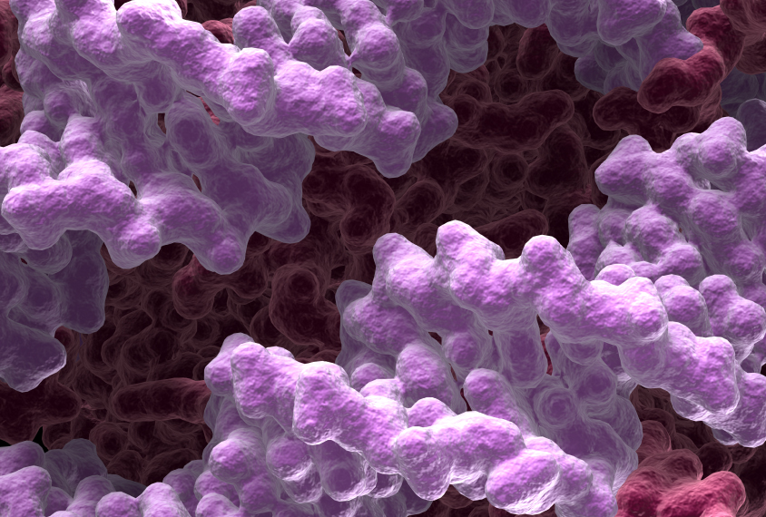
The protein spools, called histones, play a central role in controlling gene expression via the chemical tags. The tags together constitute epigenetic changes, which can alter gene expression without affecting the underlying DNA sequence.
The new study is the first to map one type of tag — an acetyl group added to histones — in autism. The researchers found that patterns of acetyl groups are more similar among autism brains than in controls.
“The most exciting thing is that we found some molecular similarity between the different [autism] brain samples,” says lead investigator Shyam Prabhakar, associate director of integrated genomics at the Genome Institute of Singapore. “Ultimately, you want to find a molecular abnormality that’s shared in most, if not all, of the individuals — and then you can start to think about how to treat it.” The work appeared 17 November in Cell.
Chromatin, or the combination of DNA and histones, is known to be altered in autism. Some people with autism have mutations in genes that regulate chromatin structure.
The addition of an acetyl group can loosen the bond between histones and DNA. This exposes DNA to the machinery that allows genes to be expressed. A study published in December by some of the same researchers found atypical patterns of gene expression in the same samples2.
“Changes in chromatin structure begin to provide a clue to the mechanisms of gene expression changes, either as causes or from compensation,” says Daniel Geschwind, professor of neurology, psychiatry and human genetics at the University of California, Los Angeles and a lead researcher on both studies.
Coil contrast:
The researchers looked at postmortem brain tissue from 45 people with autism and 49 controls. They focused on the prefrontal cortex and the temporal cortex — brain regions involved in abilities associated with autism, such as social behavior and planning. They also looked at the cerebellum, which helps to coordinate movement.
They used a protein to fish out all the histones with an acetyl tag at a site called H3K27. They then sequenced the DNA attached to the histones to create a map of the tags across the genome of each person.
They found more than 5,000 DNA regions where acetylation patterns differ between the two groups in the prefrontal cortex, and more than 7,000 such regions in the temporal cortex. By contrast, they found only 247 such regions in the cerebellum.
The companion study found that differences in gene expression between the two groups mirror this pattern, with the fewest differences in the cerebellum. This consistency of findings is “reassuring,” says Dan Arking, associate professor of genetic medicine at Johns Hopkins University in Baltimore, Maryland, who was not involved with the work. “The value of the dataset is huge,” he says.
The acetylation patterns in most of the autism brains resemble each other, suggesting a consistent autism signature. Nearly half the altered sites in the prefrontal cortex and the temporal cortex show the same pattern in each sample.
Degraded postmortem tissue can sometimes interfere with results, but histone modifications are relatively stable, says Kasper Lage, group leader at the Stanley Center for Psychiatric Research at the Broad Institute in Cambridge, Massachusetts, who was not involved in the study. This makes histones useful for analysis in postmortem brains, Lage says.
Stable signature:
The histone marks are consistent across all the autism samples, including those from a subgroup of people whose autism had a specific genetic cause. Of the 45 autism brains, 7 are from people with an extra copy of a chromosomal region called 15q11.2. The duplication leads to a condition called dup15q syndrome, characterized by severe seizures, developmental delay and autism.
Prabhakar says he expected brain tissue from individuals with dup15q syndrome to have a unique epigenetic signature. But the team found instead that the acetylation patterns in these people are similar to those seen in other forms of autism.
This sort of ‘autism signature’ may be easier to find in histone markings than in gene expression patterns, which can greatly vary between cells, says Hongjun Song, professor of neurology at Johns Hopkins School of Medicine, who was not involved with the study. “This is more reliable, in a way, than just looking at gene expression,” he says.
The researchers found an increase in acetyl groups near genes involved in brain functions that are disrupted in autism, such as signaling between neurons. The changes also cluster near some of 296 genes associated with autism. What’s more, genes near the acetylation hotspots tend to be most active around age 1, when many autism features begin to manifest.
The researchers also found roughly 2,000 DNA variants that are associated with particular acetylation changes. Two of these variants are linked to schizophrenia risk, and two others to one of five different neurological conditions, including autism. The new results suggest that the variants contribute to these conditions by altering acetyl groups.
By joining the discussion, you agree to our privacy policy.