THIS ARTICLE IS MORE THAN FIVE YEARS OLD
This article is more than five years old. Autism research — and science in general — is constantly evolving, so older articles may contain information or theories that have been reevaluated since their original publication date.
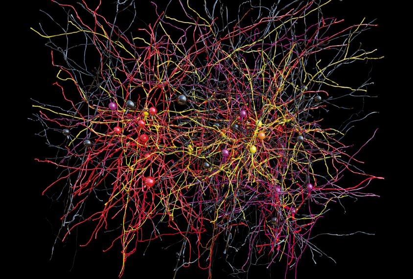
Researchers have traced the paths of thousands of neurons in a tiny piece of mouse brain, creating the largest map of neuronal wiring to date. The atlas, published in March in Nature, shows not only how these neurons connect, but also how they function as the brain processes information1.
The work is part of a massive effort, backed by more than $70 million in federal funding, to use the brain as a blueprint for intelligent machines. The findings could also offer clues about how the brain becomes wired during development and what happens when this wiring goes awry, says lead researcher R. Clay Reid, senior investigator of neural coding at the Allen Institute for Brain Science in Seattle, Washington.
“In some way that no one has thought of yet, this will help [to advance our understanding of] autism,” he says.
Previous brain-mapping efforts have focused primarily on either structure or activity. Reid’s team integrated both types of information into a single map, offering an unprecedented glimpse of the brain’s complex circuitry in action.
“This kind of fine-grained level of detail in an animal model is really important for extrapolating to understanding the human brain,” says Lucina Uddin, assistant professor of psychology at the University of Miami, who was not involved in the study. “This will help us understand some fundamental properties about neural circuits, and I think that’s a really exciting step forward.”
Grain of brain:
Reid and his team studied a mouse whose neurons glow when electrically active. They fashioned a tiny window in the mouse’s skull to watch the neurons fire as the animal looked at videos of moving geometric shapes. They used a light microscope to take pictures through the window of a piece of brain roughly the size of a grain of salt.
They then removed this tiny piece and shaved it into 3,700 ultra-thin slices to view with an electron microscope. They took roughly 10 million black-and-white pictures, which they then stitched together into one three-dimensional image. They used the branching patterns of the neurons and the surrounding blood vessels to align the images from the light and electron microscopes.
Next, the researchers traced the outlines of 1,278 neurons that send excitatory messages. They focused on the neurons that were most consistently active while the mouse viewed the videos. The 43 neurons they identified made 990 connections with other neurons.
“That’s pretty amazing, because they can actually see the interactions,” says Uddin, who scans brain structure and function in people with autism. “We can’t really ever get at that level of resolution with human brain imaging.”
Neurons that light up in response to a particular image — horizontal bars, for example — are more likely to form connections with each other than with neurons that glow in response to a different pattern, the researchers found. These connections are just as likely to span long distances as short ones, jibing with findings from a structural map released last July.
“A neuron’s function is fundamentally dependent on how it’s connected,” says Wei-Chung Allen Lee, instructor in neurobiology at Harvard University, who worked on the study. He and his colleagues used the function of the neurons as a “decoder ring” to help make sense of the complex wiring arrangements between neurons, he says.
Machine learning:
The work is a small part of a mammoth, federally funded project called MICrONS, for Machine Intelligence from Cortical Networks. Three teams of researchers from six institutions have multimillion-dollar contracts to map the structure and function of the brain’s visual center in mice and rats, with the goal of inspiring algorithms that could help driverless cars distinguish between objects such as a tree and a dog, or help computers to recognize faces.
“Even with the best super computers, visual machine learning is inferior to the basic biological visual processing in almost any animal at almost any age,” says George Church, head of synthetic biology at the Wyss Institute at Harvard and lead researcher on one of the three MICrONS projects.
Beyond helping scientists to build better computers, the findings will provide valuable insights into the basic organization of the brain. “This will help us understand the cortex,” Reid says, referring to the brain’s outer layer, which is implicated in autism. Researchers could use the new atlas of neuronal wiring to look for faulty connections in mouse models of autism.
Reid says his team will be able to map a cubic millimeter chunk of brain — roughly one-thousandth of the entire mouse brain — within five years. A third team of researchers, from Harvard University, is using a similar strategy to map a comparable piece of rat brain.
By joining the discussion, you agree to our privacy policy.